Rib Cage Anatomy Diagram - 3d Rib Cage Models Turbosquid / Rib 1 is also flattened horizontally.. Each are symmetrically paired on a right and left side. The head only articulates with the body of the t1 vertebra and therefore only one articulatory surface is present. The first rib is the widest, shortest and has the sharpest curve of all the ribs. Rib 1 is also flattened horizontally. The neck contains no bony prominences, but simply connects the head with the body.
The human rib cage is made up of 12 paired rib bones; Related posts of rib cage diagram with organs anatomy of human stomach. Rib 1 is also flattened horizontally. The first rib is the widest, shortest and has the sharpest curve of all the ribs. The human rib cage is made up of 12 paired rib bones;
Rib Cage Anatomy Human Rib Cage Info And Pictures Human Rib Cage Rib Cage Anatomy Human Ribs from i.pinimg.com Rib cage anatomy xiphoid process biology lessons human body anatomy diagram spiritual medical image. The superior surface is unique in that it is marked by two grooves that allow. The middle and upper part of your spine is called the thoracic region and it helps to support your upper body. This muscle helps rotate the upper arm. A rib has a flat body, as you can see from the picture of the anatomy of the human rib cage. The heads of ribs 1, 10, 11, and 12 have a single facet for articulation with the bodies of the thoracic vertebrae. The typical ribs have a generalised structure while the atypical ribs have variations on this structure. Gm1207196978 $ 12.00 istock in stock
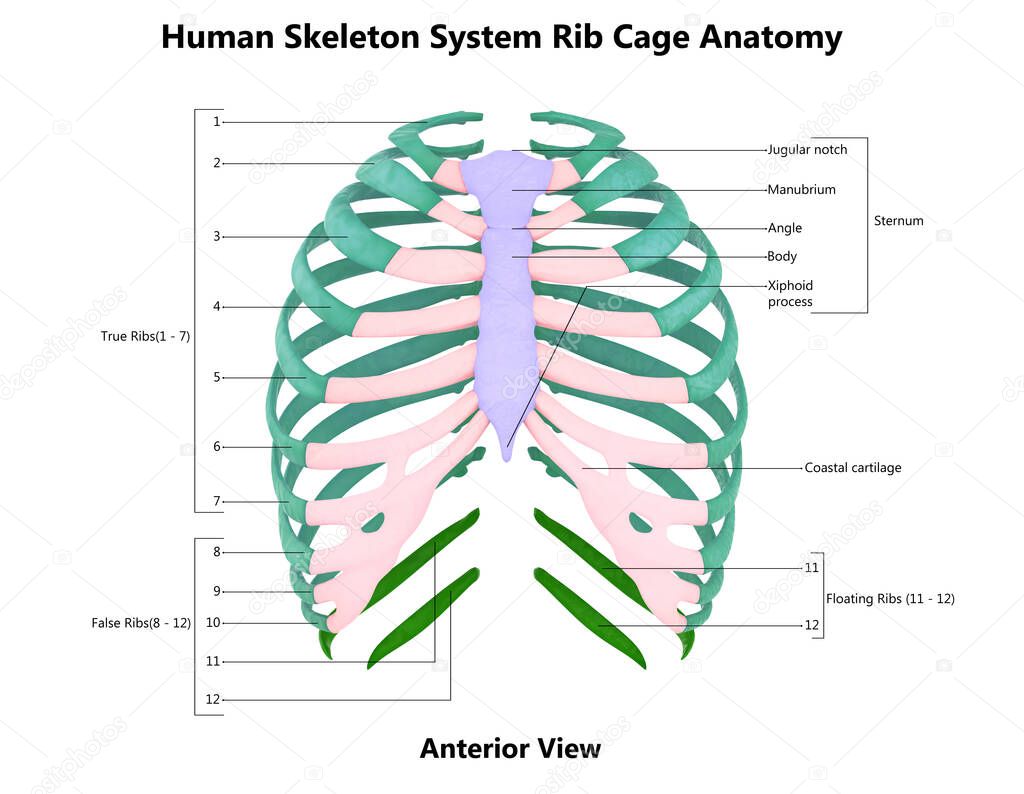
The rib cage is formed by the sternum, costal cartilage, ribs, and the bodies of the thoracic vertebrae.
The first rib is the widest, shortest and has the sharpest curve of all the ribs. A rib has a flat body, as you can see from the picture of the anatomy of the human rib cage. It also protects several vital organs of the chest, such as the heart, aorta, vena cava, and. This furrow isn't present in the 11th and 12th ribs. Our latest youtube film is ready to run. The last diagram shows how the ribs are connected to the vertebral column or spine. The heads of ribs 1, 10, 11, and 12 have a single facet for articulation with the bodies of the thoracic vertebrae. The rib cage is a bony structure found in the chest (thoracic cavity). Several muscles that move the arms, head, and neck have their origins on the sternum. Anatomy of the rib cage diagram in this image, you will find thoracic vertebrum, costochondral joint, costal cartilage, costal margin, costal arch, thoracic vertebrum, xiphoid process, xiphisternal joint, body, manubrial sternal joint, manubrium, the sternal notch in it. The typical ribs have a generalised structure while the atypical ribs have variations on this structure. Identify your right clavicle (collar bone) just below your neck (2). The rib cage, shaped in a mild cone shape and more flexible than most bone sets, is made up of varying elements such as the thoracic vertebra, 12 equally paired ribs, costal cartilage, and held together anteriorly by the sternum.
The human rib cage is made up of 12 paired rib bones; Our latest youtube film is ready to run. Rib cage pain may start in one area but travel to an area nearby. Moreover, there are many vital organs such as the heart, liver, gall bladder, kidney, and lungs under your right rib cage. The rib cage labeled diagram.
Human Skeleton System Rib Cage Bone Parts Described With Labels Anatomy Posterior View 3d 432084036 Larastock from st2.depositphotos.com Register a new.com for just $9.99 for the first year and get everything you need to make your mark online — website builder, hosting, email, and more. This muscle helps rotate the upper arm. Learn vocabulary, terms, and more with flashcards, games, and other study tools. Related posts of rib cage diagram with organs anatomy of human stomach. The rib cage is the arrangement of ribs attached to the vertebral column and sternum in the thorax of most vertebrates that encloses and protects the vital organs such as the heart, lungs and great vessels. The rib cage, shaped in a mild cone shape and more flexible than most bone sets, is made up of varying elements such as the thoracic vertebra, 12 equally paired ribs, costal cartilage, and held together anteriorly by the sternum. Learn vocabulary, terms and more with flashcards, games and other study tools. The first rib is the widest, shortest and has the sharpest curve of all the ribs.
The superior surface is unique in that it is marked by two grooves that allow.
Rib cage anatomy xiphoid process biology lessons human body anatomy diagram spiritual medical image. The superior surface is unique in that it is marked by two grooves that allow. Gm1207196978 $ 12.00 istock in stock The last diagram shows how the ribs are connected to the vertebral column or spine. This furrow isn't present in the 11th and 12th ribs. The typical rib consists of a head neck and body. The bones of the rib cage are the sternum, the 12 thoracic vertebrae and the 12 pairs of ribs. Rib cage anatomy, labeled vector illustration diagram. The cartilage that forms at the end of each rib (costal cartilage) attaches either. The human rib cage is made up of 12 paired rib bones; Animated full human body anatomy. Located in the rib cage, this muscle keeps the shoulder blade against the chest wall and helps rotate the shoulder blade. The rib cage, shaped in a mild cone shape and more flexible than most bone sets, is made up of varying elements such as the thoracic vertebra, 12 equally paired ribs, costal cartilage, and held together anteriorly by the sternum.
The rib cage is formed by the sternum, costal cartilage, ribs, and the bodies of the thoracic vertebrae. The front edge ends with an ellipsoidal shape on which. Pork ribs the linchpin of the texas trinity by daniel vaughn. Anatomy of human stomach 10 photos of the anatomy of human stomach anatomy human colon, anatomy human digestive system, anatomy human heart, anatomy human kidney, anatomy human liver, anatomy human pancreas, anatomy human spleen, human body stomach, stomach, anatomy human colon, anatomy human digestive system, anatomy. Anatomy of the rib cage diagram in this image, you will find thoracic vertebrum, costochondral joint, costal cartilage, costal margin, costal arch, thoracic vertebrum, xiphoid process, xiphisternal joint, body, manubrial sternal joint, manubrium, the sternal notch in it.
Thoracic Rib Cage Anatomy In Detail Anterior View from www.anatomynote.com The typical rib consists of a head, neck and body: This furrow isn't present in the 11th and 12th ribs. Ribs 11 and 12 do not have necks or tubercles and the anterior tips of. Gm1207196978 $ 12.00 istock in stock Anatomy of human stomach 10 photos of the anatomy of human stomach anatomy human colon, anatomy human digestive system, anatomy human heart, anatomy human kidney, anatomy human liver, anatomy human pancreas, anatomy human spleen, human body stomach, stomach, anatomy human colon, anatomy human digestive system, anatomy. Rib cage pain may start in one area but travel to an area nearby. Über 7 millionen englischsprachige bücher. The rib cage, shaped in a mild cone shape and more flexible than most bone sets, is made up of varying elements such as the thoracic vertebra, 12 equally paired ribs, costal cartilage, and held together anteriorly by the sternum.
The human rib cage is made up of 12 paired rib bones;
Related posts of rib cage diagram with organs anatomy of human stomach. The sternum is a flat bone that is made up of three parts, the (1) manubrium, (2) body, and the (3) xiphoid process. The front edge ends with an ellipsoidal shape on which. Anatomy of the rib cage diagram in this image, you will find thoracic vertebrum, costochondral joint, costal cartilage, costal margin, costal arch, thoracic vertebrum, xiphoid process, xiphisternal joint, body, manubrial sternal joint, manubrium, the sternal notch in it. Anatomy of human stomach 10 photos of the anatomy of human stomach anatomy human colon, anatomy human digestive system, anatomy human heart, anatomy human kidney, anatomy human liver, anatomy human pancreas, anatomy human spleen, human body stomach, stomach, anatomy human colon, anatomy human digestive system, anatomy. Identify the costal margin (lower border of the rib cage) on your right side. Diagram rib cage with organs / thoracic cavity description anatomy physiology britannica. The point of intersection between an imaginary line drawn from the middle of the right clavicle downwards and the right costal margin can be identified (2). The rib cage is a bony structure found in the chest (thoracic cavity). This is same as the tip of 9th costal. These are cut from the longer straight portion of the rib cage below the baby backs. Each pair is numbered based on their attachment to the sternum, a bony process at the front of the rib cage which serves as an anchor point. This furrow isn't present in the 11th and 12th ribs.
Rib cage anatomy, labeled vector illustration diagram rib cage anatomy. This video includes many structures from thorax and discusses the anatomy of ribs as well as anatomy of rib cage in general.


0 Komentar